Cytoplasmic Protein Imaging with Mid-Infrared Photothermal Microscopy: Cellular Dynamics of Live Neurons and Oligodendrocytes
| author | Minhaeng Cho |
|---|---|
| Homepage | https://cmsd.ibs.re.kr/html/cmsd_en/ |
| journal | J. Phys. Chem. Lett. 2019, 10, 2857−2861 |

Chanjong Park(통합과정, 제1저자)
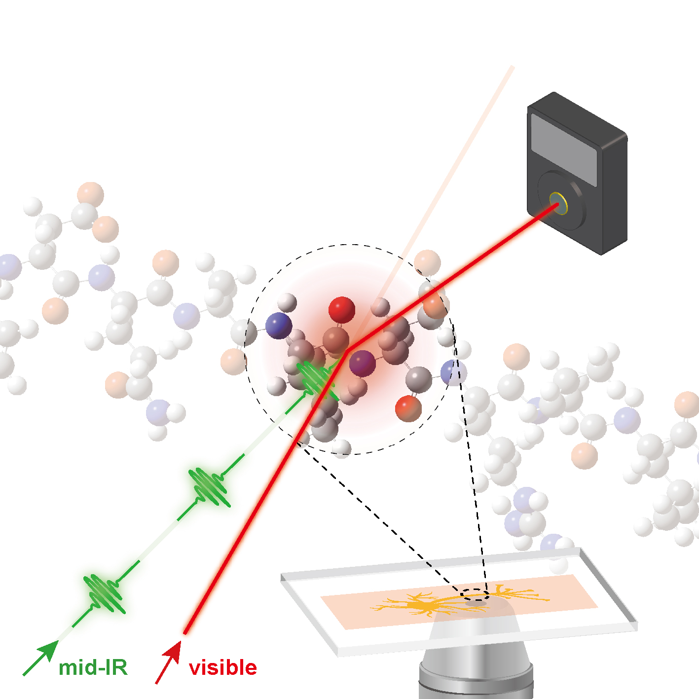
Mid-infrared photothermal microscopy has been suggested as an alternative to conventional infrared microscopy because in addition to the inherent chemical contrast available upon vibrational excitation, it can feasibly achieve spatial resolution at the submicrometer level. Furthermore, it has substantial potential for real-time bioimaging for the observation of cellular dynamics without photodamage or photobleaching of fluorescent labels. We performed real-time imaging of oligodendrocytes to investigate cellular dynamics throughout the life cycle of a cell, revealing details of cell division and apoptosis, as well as cellular migration. In the case of live neurons, we observed a photothermal contrast associated with traveling protein complexes on an axon, which correspond to the transport of vesicles from the cell body to the dendritic branches of the neuron through the cytoskeleton. We anticipate that mid-infrared photothermal imaging will be of great use for gaining insights into the field of biophysical science, especially with regard to cellular dynamics and functions.
https://pubs.acs.org/doi/10.1021/acs.jpclett.9b00616
« Prev Structure–thermopower relationships in molecular thermoelectrics
 Structure–thermopower relationships in molecular thermoelectrics
2019.06.07by webmaster
〈
Structure–thermopower relationships in molecular thermoelectrics
2019.06.07by webmaster
〈
Conjugated Polyelectrolytes as Multifunctional Passivating an... Next »
 Conjugated Polyelectrolytes as Multifunctional Passivating an...
2019.06.07by webmaster
〉
Conjugated Polyelectrolytes as Multifunctional Passivating an...
2019.06.07by webmaster
〉
Articles
-
 Significantly Improved Morphology and Efficiency of Nonhalogenated Solvent-Proces...
-
 Mechanical Force Induces Ylide-Free Cycloaddition of Nonscissible Aziridines
-
 Bright ligand-activatable fluorescent protein for high-quality multicolor live-ce...
-
 Mid-wavelength Infrared Photoluminescence and Lasing of Tellurium Element Solid a...
-
 Power Factor of One Molecule Thick Films and Length Dependence
-
 An Emerging Molecular Design Approach to Heavy-Atom-Free Photosensitizers for Enh...
-
 Ga‐Based Liquid Metal Micro/Nanoparticles: Recent Advances and Applications
-
 A Hydrogen-Bonded Organic Framework with Type IV NH3 Adsorption Behavior
-
 Janus to Core-Shell to Janus: Facile Cation Movement in Cu2-xS/Ag2S Hexagonal Nan...
-
 Two Different Length-Dependent Regimes in Thermoelectric Large-Area Junctions of ...
-
 From p-Xylene to Ibuprofen in Flow: 3-Step Synthesis via Unified Sequence of Chem...
-
 Mid-wavelength Infrared Photoluminescence and Lasing of Tellurium Element Solid a...
-
Structure–thermopower relationships in molecular thermoelectrics
-
 Cytoplasmic Protein Imaging with Mid-Infrared Photothermal Microscopy: Cellular D...
-
Conjugated Polyelectrolytes as Multifunctional Passivating and Hole‐Transporting ...
-
 Covalently Linked Perylene Diimide–Polydiacetylene Nanofibers Display Enhanced St...
-
 Blue Emission of α-GaN Colloidal Quantum Dots via Zn Doping
-
 Gas‐phase conformations of intrinsically disordered proteins and their complexes ...
-
 Multifunctional Self-Doped Nanocrystal Thin-Film Transistor Sensors
-
 Fine-tuning of wettability in a single metal–organic framework via postcoordinati...
Designed by sketchbooks.co.kr / sketchbook5 board skin